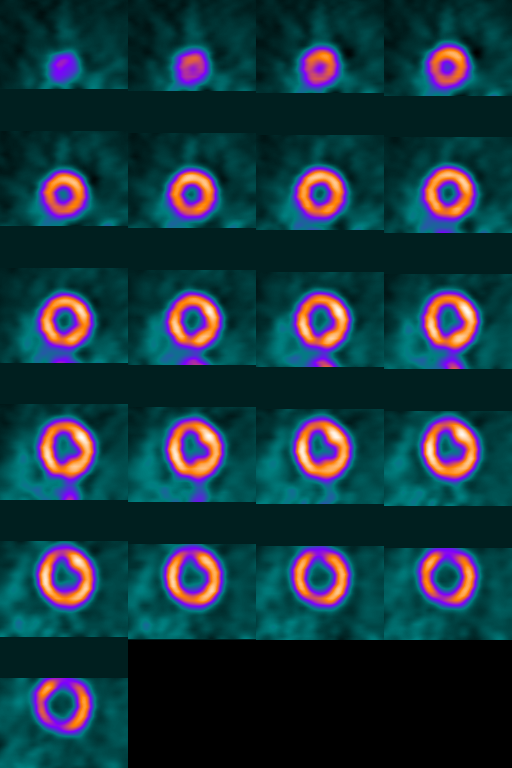
Volume Slicer Applet - PET
This is a scan of a normal human left ventricle taken by the
Positron Emission Tomography (PET) scanner at Crawford-Long
Hospital of Emory University. The patient was injected with a
blood-flow tracing agent (Rb82) that is a positron emitter.
The dataset is made up of 21 slices, each 128x128.
The voxels are sampled at 1.275x1.275x4.505 mm by the scanner.
The pixels of the ZY and XZ planes are interpolated to
1.275x1.275 mm to make these slices isotropic (interpolation
artifacts are quite clear).
| XY Plane |
ZY Plane |
|
|
|
 |
|
 |
| XZ Plane |
|
|
| Position cursor over any
of the slices. The slice is highlighted and color-coded hashmarks
are highlighted in the other viewers showing the position of the
active slice. Use the 'j' and 'k' keys to move through the volume
(up/down and space/bs work too).
|
You are looking at the gif version of this volume (163 KB).
If your browser can handle jpeg images in an applet (Netscape for
HP doesn't), then you can look at the
jpeg version (37 KB).
You really need a good 24-bit color monitor to get the full color range.
Created 12/04/95 by abb@nuccard.eushc.org
Updated 3/31/96